Corneal Ulcer Classification in Veterinary Care
Aug 19, 2025

Have you ever wondered why your pet suddenly starts pawing at their eye?
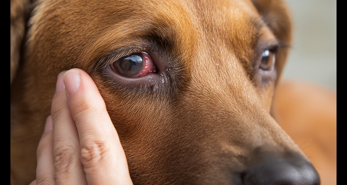
When a pet starts squinting, tearing, or pawing at their eye, it can make the pet owners very worried. One possible cause is a corneal ulcer, which is a wound on the clear surface of the eye. However, not all ulcers are the same. Knowing the corneal ulcer classification is important for proper diagnosis and treatment plan.
For veterinarians, this knowledge isn’t just about medicine. It can be the difference between saving an eye and losing it. For pet owners, understanding the types of corneal ulcers can lead to getting help before permanent damage occurs.
In this article, we will discover six main types of corneal ulcers in dogs. We will explain how they differ and connect every kind to its suggested treatment. Whether you’re getting ready for veterinary CE or want to protect your pet’s vision, knowing the corneal ulcer classification is very important.
1. Superficial Corneal Ulcer
This is the mildest form, affecting only the thin outer layer of the cornea. The edges are usually clear, and the ulcer can appear anywhere on the cornea. If the lesion goes under the third eyelid, veterinarians look for a hidden foreign body.
Superficial ulcers heal with medical treatment, usually within a week. They can impact dogs of any breed or age and are seen as uncomplicated unless an infection occurs.
Although a superficial ulcer may seem normal, some types do not heal with drops alone. This is when we encounter the more stubborn detached-edge ulcer.
2. Detached-Edge Ulcer (SCCED)
Also called an indolent or Boxer ulcer, this type is always superficial and has loose, poorly attached edges. Under fluorescein staining, the dye spreads beneath the edges. This ulcer often occurs in middle-aged dogs and is not known for healing with drops alone.
The current best treatment is a diamond burr debridement, which encourages new healthy cells to attach to the cornea. Without this, the ulcer may last for weeks.
If SCCED is persistent but stays limited to the surface, the following type, stromal corneal ulcer, occurs when the damage goes deep.
3. Stromal Corneal Ulcer
This ulcer goes into the stroma, which is the thick middle layer of the cornea. The damage can either be shallow or deep, but the edges are typically sharp and clear. The fluorescein dye can move beyond the edges because it spreads through the layers of corneal collagen.
Treatment usually begins with strong medical therapy, but surgery may be necessary if the ulcer is deep or infected. It is highly recommended to use cytology and culture to help choose the right antibiotic.
When the stromal tissue is completely gone, only one delicate layer remains between the outside world and the inside eye. This is the dangerous descemetocele stage.
4. Descemetocele
Here, the corneal stroma is entirely absent. It leaves only the Descemet’s membrane, which is a fragile and transparent layer. This ulcer condition in dogs often looks gelatinous in the center and should be treated as a clinical emergency.
In Brachycephalic breeds, descemetoceles usually occur in the center. Surgical repair must happen quickly to prevent rupture. Cultures and cytology are best taken while the animal is under anesthesia for surgery.
Unfortunately, if this thin layer ruptures, it results in a full-thickness wound.
5. Perforated Corneal Ulcer
This happens when all layers of the cornea are missing. The defect may not be easy to see because fibrin or iris tissue can cover the opening. In severe cases, there may be fluid leaking from the front chamber, which can be confirmed with a Seidel test.
Flat-faced breeds are more likely to have central perforations. Quick surgery is needed to restore the globe’s structure, and vets should collect diagnostic samples before the repair.
There is also a type of corneal ulcer that can quickly damage the cornea.
6. Corneal Melting Ulcer
Also known as Keratomalacia, this condition is caused by enzymes known as collagenases that dissolve the central stroma. The cornea appears soft and gelatinous, and the edges lose their sharpness.
This is a real emergency. Immediate medical treatment is necessary. Sometimes, corneal cross-linking is done to strengthen the tissue. Surgery may be required if the damage worsens.
Recognizing the destructive potential of ulcers underscores the importance of each type. This is crucial for a pet’s vision.
Also Read: “Is it urgent that we seek an ophthalmologist…”
Why Understanding The Corneal Ulcer Classification Matters
Each ulcer requires its own approach. A superficial abrasion might heal with eye drops. In contrast, a melting ulcer needs intensive hospital care. Early recognition and correct classification increase the chances of saving vision and reducing pain.
For veterinarians who want to improve their diagnostic skills, the corneal ulcers webinar for veterinary professionals offers real-world case studies and effective management strategies. You’ll learn how to diagnose and treat confidently, backed by the latest research and clinical insights.
If you prefer a quick learning format during the day, Lunchtime CE is the way to go. It provides brief, valuable updates in short, engaging sessions. Each session offers practical takeaways that can be used right away in practice, without needing to take time away from patient care.
Final Thoughts
The eyes are delicate. A type of corneal ulcer can mean the difference between a quick recovery and lasting damage. By understanding the different types of corneal ulcers, from simple scratches to serious perforations, pet owners can act quickly, and veterinarians can pick the best treatment.
If your pet shows any signs of eye pain, don’t wait. What seems minor today could turn into an emergency tomorrow. The right knowledge, used at the right time, can protect your pet’s vision for life. Additionally, lunchtime CE for veterinarians is perfect for their busy schedules. Ongoing education through veterinary CE is the best way to keep those eyes bright and healthy.
FAQs
Q: What are the classifications of corneal ulcers?
Corneal ulcers are usually divided into six main types: superficial, detached-edge (also known as SCCED), stromal, descemetocele, perforated, and melting ulcers. Each type varies in depth, severity, and urgency. This classification helps veterinarians select the best treatment and estimate healing time.
Q: What are the different types of corneal ulcers in dogs?
Dogs can develop all six types of corneal ulcers, ranging from mild superficial lesions to severe melting ulcers. Some heal quickly with topical medications. Others, particularly descemetoceles and perforated ulcers, need urgent surgery. Identifying the type early can determine whether a dog fully recovers or suffers permanent vision loss.
Q: What is the difference between complicated and uncomplicated corneal ulcers?
Uncomplicated ulcers are shallow, non-infected, and typically heal within 1-2 weeks with basic medical treatments. Complicated ulcers are deeper, infected, slow-healing, or recurrent, and they often need more advanced treatments or surgery. Quick evaluation by a vet helps ensure that a simple ulcer doesn’t complicate.


Disclaimer: healthcareforpets.com and its team of veterinarians and clinicians do not endorse any products, services, or recommended advice. All advice presented by our veterinarians, clinicians, tools, resources, etc is not meant to replace a regular physical exam and consultation with your primary veterinarian or other clinicians. We always encourage you to seek medical advice from your regular veterinarian.

